Skeletal muscle under microscope with labeled diagram » anatomylearner Muscle skeletal structure myofilaments myofibril lab contractile proteins Muscle skeletal microscopic anatomy tissue chap11
Skeletal Muscle Under Microscope
Microscopic muscle anatomy anatomy diagram book
Skeletal muscle under microscope
Muscle microscopic skeletal anatomy muscular systemSkeletal muscle fiber structure Skeletal tissue labeled copied permission physiology membrane attribution licenseContraction in the simplest sense is shortening of a muscle fibre. when.
Microscopic structure of skeletal muscle fibers diagramSkeletal structure fibre contraction muscular physiology steemit gross macro Muscle cell labeled muscular system anatomy human anatomy andThe muscular system.

Anatomy: chapter 7: macroscopic muscle anatomy (diagram!!) diagram
900 skeletal muscle microscope images, stock photos, 3d objectsMicroscopic anatomy of skeletal muscle youtube Skeletal muscle is characterized by its cylindrical shape. it isHuman structure virtual microscopy.
Muscular system anatomy and physiologyDescribe the microscopic features of osseous tissue Muscular system anatomy physiology nurseslabs muscle fibers body cells gross called myosin made nursing some school exercise human also study6.2 quiz: microscopic anatomy of skeletal muscles.

Muscle skeletal microscopic structure
Microscopic structure of skeletal muscle by dr. s. n. singhMacroscopic and microscopic structure of muscle tissue Schematic relating the biochemical components to the microscopic(a) illustration of skeletal muscle structure copied with permission.
Structure of skeletal muscle dissection of the skeletal muscle bellyDraw a neat labelled diagram of striated muscle labeled diagram [diagram] labelled diagram of skeletal muscle tissueCardiac muscle slides.

Describe the macroscopic and microscopic anatomy of a skeletal muscle
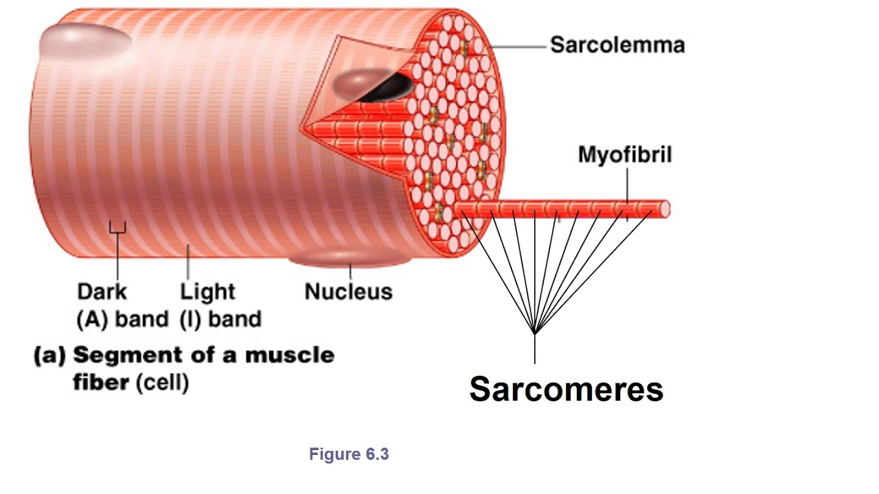
Structure of skeletal muscle – earth's labSkeletal muscle: structure and contraction Muscle cardiac striated types between skeletal histology cells intercalated discs structure fibers slides human striations differences nuclei located microscopy featuresSkeletal fiber sarcolemma membrane cytoplasm called sarcoplasm myofibrils fibers plasma contraction surrounded appearance banded fibrils composed.
.